

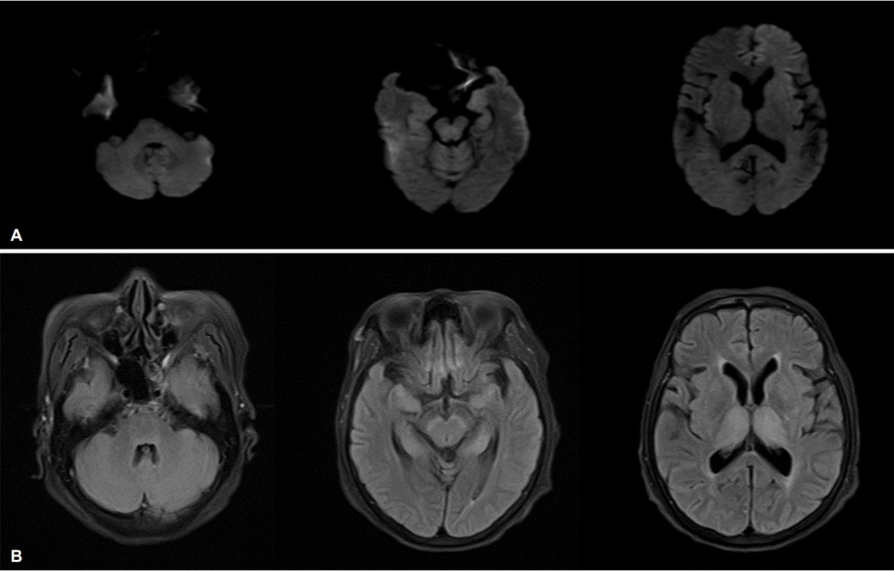
68세 여자가 혼동과 고열(39℃)로 내원하였다. 증상 발생 후 1일째에 시행한 뇌 자기공명영상에서는 특별한 이상 소견은 보이지 않았고, 뇌척수액검사에서 백혈구(100/μL, lymphocyte 53%), 단백질(91.8 mg/dL) 및 adenosine deaminase (21 U/L)의 증가 소견을 보였다. 환자의 의식수준은 점차 혼수상태로 악화되었으며 6일 뒤에 시행한 자기공명영상에서 대칭적인 고음영의 병터가 양쪽 뇌줄기, 다리뇌수 뇌연결 피개 부위, 흑색질, 해마, 시상에서 관찰되었다(Fig.). 특징적인 영상 소견을 토대로 시행한 항체검사에서 일본뇌염이 진단되었다(혈청 immunoglobulin M [IgM] 역가: 1:16→1:128 (10일 뒤), 뇌척수액 IgM 역가: 40.9 immune status ratio). 대부분의 뇌염 환자에서 자기공명영상검사는 발생 수일 내에 시행되는데, 이 시기에서는 일본뇌염의 특징적인 이상이 나타나지 않을 수 있으며, 수일 뒤에 지연되어 나타날 수 있다[1,2].
| J Korean Neurol Assoc > Volume 37(2); 2019 > Article |

|
REFERENCES
1. Handique SK, Das RR, Barman K, Medhi N, Saharia B, Saikia P, et al. Temporal lobe involvement in Japanese encephalitis: problems in differential diagnosis. AJNR Am J Neuroradiol 2006;27:1027-1031.

2. Kalita J, Misra UK, Mani VE, Bhoi SK. Can we differentiate between herpes simplex encephalitis and Japanese encephalitis? J Neurol Sci 2016;366:110-115.

Figure.
(A) The initial diffusion weighted MRI of the brain performed one day after symptom onset demonstrates no abnormal lesions. (B) The follow-up MRI 6 days later demonstrated symmetric high signal intensities in the periaqueductal areas of the brainstem, pontomedullary junction tegmentum, bilateral substantia nigra, hippocampus, and thalami on fluid-attenuated inversion recovery images. MRI; magnetic resonance imaging.
- TOOLS
 PDF Links
 PubReader
 ePub Link
 Full text via DOI
 Download Citation
 Print
Print


-
METRICS

-
- 0 Crossref
- 0 Scopus
- 2,915 View
- 126 Download
-

- Related articles
-
Magnetic Resonance Image Findings in Venous Compression of Trigeminal Nerve2021 May;39(2)
Carotid Web: Ultrasonographic and Magnetic Resonance Image Findings2018 August;36(3)
In Vivo Proton Magnetic Resonance Spectroscopic Findings in Brain Abscess: A Case Report2007 ;25(3)
Temporal Bone Magnetic Resonance Imaging Study in Hemifacial Spasm2000 ;18(3)
- Editorial Office
-
(ZIP 03163) #1111, Daeil Bldg, 12, Insadong-gil, Jongno-gu, Seoul, Korea
Tel: +82-2-737-6530 Fax: +82-2-737-6531 E-mail: jkna@neuro.or.kr
Copyright © 2024 by Korean Neurological Association.

