

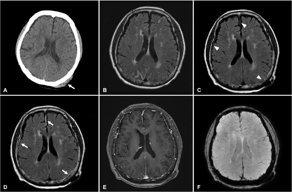
75세 여자가 척추관협착증으로 꼬리차단마취 후 가역적뇌혈관수축증후군 진단 하에 경과 관찰 중 침대에서 낙상하였고 당시에 의식소실 및 신경계 장애는 없었으나 좌측 후두부 부종이 관찰되었다. 뇌 컴퓨터단층검사에서 좌측 후두부 두피하 출혈 외에 이상 소견은 없었다(Fig. A). 낙상 후 3일째 시행한 액체감쇠역전회복영상에서 우측 전두엽, 좌측 후두엽과 대뇌반구 사이 뇌막에서 미세한 고신호강도 병변이 관찰되었고 이 병변은 뚜렷하게 조영증강되었으나 동시에 시행된 자화강조영상과 조영증강 T1강조영상 및 낙상 이전에 시행한 조영증강 액체감쇠역전회복영상에서는 명확한 이상이 관찰되지 않았다(Fig. B-F). 외상성 뇌 손상 후 조영증강 액체감쇠역전회복영상에서 뇌막의 조영증강 현상은 다른 영상검사에 비하여 더 흔하게 관찰되는 이상 소견으로 보고되었다[1,2]. 조영증강 액체감쇠역전회복영상은 일반적으로 시행되는 자기공명영상 기법은 아니지만 경미한 두부 외상에서 진단적 유용성이 있을 것으로 생각된다.
| J Korean Neurol Assoc > Volume 37(1); 2019 > Article |

|
REFERENCES
1. Chiara Ricciardi M, Bokkers RP, Butman JA, Hammoud DA, Pham DL, Warach S, et al. Trauma-specific brain abnormalities in suspected mild traumatic brain injury patients identified in the first 48 hours after injury: a blinded magnetic resonance imaging comparative study including suspected acute minor stroke patients. J Neurotrauma 2017;34:23-30.

2. Kim SC, Park SW, Ryoo I, Jung SC, Yun TJ, Choi SH, et al. Contrast-enhanced FLAIR (fluid-attenuated inversion recovery) for evaluating mild traumatic brain injury. PLoS One 2014;9:e102229.
Figure 1.
Brain CT and magnetic resonance images. (A) Brain CT shows no intracranial hemorrhage and skull fracture. Scalp hematoma (arrow) on left occipital area is noted. (B) There is no meningeal lesion on Gadolinium enhanced FLAIR image before the head trauma. (C, D) There are subtle high signal intensity meningeal lesions on FLAIR (arrowheads). Gadolinium enhanced FLAIR image shows meningeal enhancement on the left frontal, right occipital and interhemispheric fissure (arrows). (E, F) Contrast-enhanced T1 weighted image and susceptibility weighted image show no abnormal enhancement and hemorrhage. CT; computed tomography, FLAIR; fluid attenuated inversion recovery.
- TOOLS
 PDF Links
 PubReader
 ePub Link
 Full text via DOI
 Download Citation
 Print
Print


-
METRICS

-
- 0 Crossref
- 0 Scopus
- 3,513 View
- 59 Download
-

- Related articles
- Editorial Office
-
(ZIP 03163) #1111, Daeil Bldg, 12, Insadong-gil, Jongno-gu, Seoul, Korea
Tel: +82-2-737-6530 Fax: +82-2-737-6531 E-mail: jkna@neuro.or.kr
Copyright © 2024 by Korean Neurological Association.

